
The Basics of Electromyography and Its Clinical Uses
Electromyography (EMG) is a diagnostic technique used to assess the electrical activity of muscles and the nerves that control them. This test provides critical insights into neuromuscular health, aiding in the diagnosis of a wide range of conditions, from nerve injuries to degenerative muscle diseases. Initially developed in the early 20th century, EMG has since evolved into a sophisticated tool that integrates advanced signal processing and artificial intelligence, improving diagnostic precision.
Understanding Electromyography
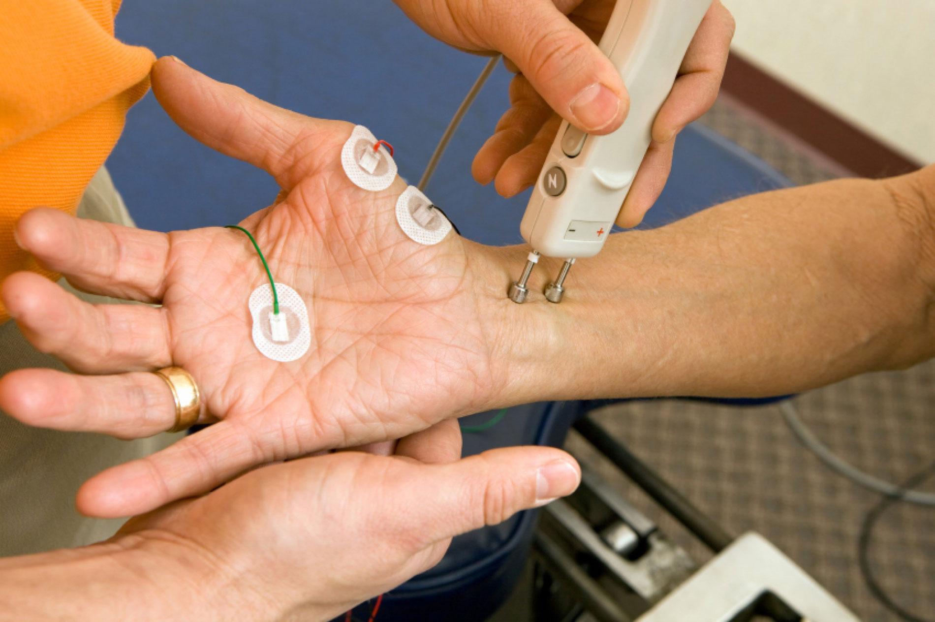
EMG measures the electrical signals produced when muscle fibers contract. The test can be performed using two primary methods: surface EMG (sEMG) and needle EMG. Surface EMG involves placing electrodes on the skin, ideal for general muscle activity assessment. In contrast, needle EMG involves inserting fine electrodes directly into the muscle, offering more precise data on deeper neuromuscular function. The electrical signals captured during EMG help physicians distinguish between healthy muscle activity and pathological conditions.
The Science Behind EMG Signals
Muscle contractions are governed by motor neurons, which transmit electrical impulses from the brain and spinal cord to the muscles. When a nerve signal reaches a muscle fiber, it triggers an action potential, leading to contraction. A well-functioning neuromuscular system exhibits a distinct pattern of electrical activity. Conversely, abnormal EMG readings—such as excessive spontaneous activity or irregular signal transmission—may indicate underlying nerve damage or muscle disease.
The EMG Procedure: What to Expect
Before an EMG test, patients may be advised to avoid caffeine and certain medications that could influence muscle activity. During the procedure, electrodes are either attached to the skin or inserted into the muscle. The patient may be asked to contract or relax specific muscles while the machine records electrical responses. The test may cause mild discomfort, particularly with needle EMG, but it is generally well-tolerated. The entire process typically lasts between 30 and 60 minutes.
Clinical Applications of Electromyography
EMG is an essential tool for diagnosing and monitoring neuromuscular disorders. It helps physicians identify peripheral nerve injuries, such as carpal tunnel syndrome and assess muscle diseases, including polymyositis and myasthenia gravis. EMG also plays a crucial role in understanding unexplained muscle weakness, spasms and chronic pain conditions.
EMG in Neurological Disorders
Certain neurological conditions manifest with distinct EMG abnormalities. For instance, amyotrophic lateral sclerosis (ALS) presents with characteristic spontaneous muscle activity, while muscular dystrophy exhibits altered action potentials. In cases of multiple sclerosis (MS), EMG can aid in evaluating nerve signal transmission impairments. Additionally, spinal cord injuries can be assessed through EMG, offering crucial insights into nerve regeneration potential.
EMG in Orthopedic and Rehabilitation Medicine
In orthopedic and rehabilitation settings, EMG is invaluable in post-surgical recovery assessments. By analyzing muscle function after procedures like knee replacements or spinal surgeries, clinicians can tailor rehabilitation strategies. EMG is also used to optimize physical therapy programs, ensuring targeted muscle engagement. Moreover, advancements in prosthetic technology integrate EMG signals to improve the responsiveness of artificial limbs.
Advancements in EMG Technology
The field of electromyography has seen remarkable technological progress. High-density EMG (HD-EMG) offers detailed spatial resolution of muscle activity, enhancing diagnostic precision. The emergence of wireless and wearable EMG devices has enabled real-time monitoring, particularly beneficial in sports science and rehabilitation. Additionally, artificial intelligence (AI) and machine learning algorithms are revolutionizing EMG interpretation, reducing human error and improving diagnostic accuracy.
Limitations and Challenges of EMG
Despite its diagnostic utility, EMG has limitations. The test’s accuracy can be influenced by patient-related factors, such as anxiety-induced muscle tension or movement artifacts. Additionally, false-positive and false-negative results remain a concern, necessitating corroborative diagnostic tests like nerve conduction studies. Researchers continue to explore ways to improve EMG reliability, including enhanced signal processing techniques and automation.
Conclusion
Electromyography remains a cornerstone of neuromuscular diagnostics, helping clinicians detect, monitor and manage a wide range of conditions. As technology advances, EMG continues to evolve, integrating with AI-driven analysis and non-invasive monitoring tools. Its role in clinical medicine is poised to expand, offering deeper insights into muscle and nerve function while improving patient outcomes.
